Product details
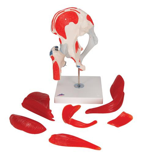
Full-size, normal right hip with proximal femur and lower lumbar vertebrae including sciatic nerve, gluteus medius, gluteus minimus, iliacus, inferior and superior gemellus, obturator internus, pyriformis and psoas muscles, articular capsule ligaments, and L4-L5 with sacrum. Includes
6-1/4 in. x 8-1/4 in. information card. Model size: 7-1/2 in. x 4-3/4 in. x 10 in. Base size: 8-3/16 in. x 6-1/4 in.
The Muscled Hip with Sciatic Nerve Model (SB49530) offers a detailed anatomical representation of the hip region, highlighting major muscles and the sciatic nerve. This model provides clear visualization of muscular structures and nerve pathways essential for understanding hip function and related conditions.
Perfect for medical students, physical therapists, and healthcare professionals, it serves as an effective tool for teaching anatomy, diagnosing sciatic nerve issues, and planning treatments. The durable design is suitable for frequent use in educational and clinical settings.
Features:
-
Detailed anatomy of hip muscles and sciatic nerve
-
Highlights muscle attachments and nerve pathways
-
Ideal for medical education, physical therapy, and clinical training
-
Durable and easy to handle for repeated demonstrations
-
Enhances understanding of hip mechanics and nerve-related conditions

