Product details
- healthy
- malignant cells are found at the surface, within the epidermis
- malignant cells fill the epidermis, a few invade the papillary layer
- malignant cells fill the papillary layer
- malignant cells invade the reticular layer
- malignant cells have reached the subcutaneous fatty tissue, satellite cells approach a vein
The skin cancer model is a great tool for illustrating this skin pathology.
Every original 3B Scientific® Anatomy Model gives you direct access to its digital twin on your smartphone, tablet or desktop device.
Enjoy using the exclusive virtual anatomy content with the following features:
- Freely rotate your digital model and zoom in and out
- Display hotspots and their anatomical structures
- Augmented Reality (AR) feature starts your virtual anatomy model
- Anatomy Quiz function to test and improve your anatomical knowledge with instant results and final score evaluation
- Drawing function that allows image customization with save and share function
- Useful Notes function to help you with your personal learning
- Possibility to learn both male and female anatomy
- Easy access to 3D content both online and offline
- Available in 11 languages
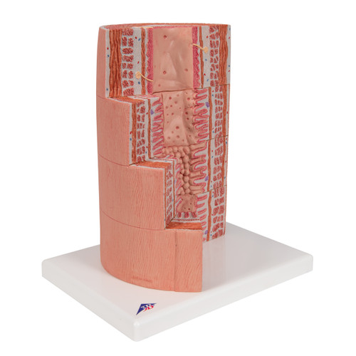
Educate with confidence using a model that removes ambiguity from difficult conversations about skin cancer. This cross-section of human skin is enlarged eightfold to make micro-anatomy readable at a glance: epidermis, dermis, and hypodermis, plus accessory structures such as hair follicle, sebaceous gland, and sweat gland. Side-by-side panels (or zones) illustrate five milestones in melanoma development, helping learners compare normal and pathological appearances in one view.
The sequence typically depicts:
-
Normal skin with intact layers and adnexa.
-
Atypical/dysplastic changes (abnormal melanocyte proliferation).
-
Melanoma in situ / radial growth within the epidermis.
-
Vertical growth phase with dermal invasion.
-
Advanced melanoma with deeper tissue involvement and potential vascular/lymphatic spread.
Each stage is sculpted with realistic contours and color transitions so you can point to borders, asymmetry, and depth—key factors behind the ABCDEs and Breslow thickness discussion. The stable base keeps the model upright for hands-on demonstrations, while the included 3B Smart Anatomy access provides supplemental digital content to reinforce learning.
Use it to:
-
Explain biopsy results and treatment rationales.
-
Demonstrate why early detection matters.
-
Train students on staging concepts and terminology.
-
Support community outreach, health fairs, and melanoma awareness events.
Note: Educational device only. Not a diagnostic tool.
Key Features
-
Five-stage melanoma progression shown in one model for instant comparison.
-
8× magnification reveals layer boundaries, lesion depth, and tissue changes.
-
High-contrast coloring & tactile detail aid group teaching and patient counseling.
-
Durable, medical-grade construction on a stable desktop base; easy to sanitize.
-
3B Smart Anatomy access for interactive digital learning (mobile & web).
-
Compact footprint fits exam rooms, sim labs, and lecture podiums.
What’s Included
-
Skin Cancer Model with 5 stages, mounted on base
-
Printed reference/overview card (labels & stage summaries)
-
3B Smart Anatomy activation (digital content access)
Specifications (Approx.)
-
Magnification: 8× cross-section
-
Stages Displayed: 5 (from normal skin to advanced melanoma)
-
Format: Desktop, base-mounted teaching model
-
Materials: Rigid, wipe-clean polymer; matte, glare-reduced finish
-
Brand / Program: 3B Smart Anatomy
-
SKU: 005-1000293
(Exact dimensions/weights may vary slightly by production lot.)
Who It’s For
-
Dermatology & Oncology clinics (patient education)
-
Family practice & urgent care (early-detection counseling)
-
Nursing, PA, and medical schools (staging & pathology teaching)
-
Public health educators, community screening & awareness programs
-
Aesthetic/skin-care practices emphasizing sun safety & lesion monitoring
Care & Cleaning
Wipe with a soft cloth dampened with mild soap and water or 70% isopropyl alcohol. Avoid abrasive pads, strong solvents, and prolonged direct sunlight.
| Weight | 0.75 lb |
| Dimensions | 5.5 x 3.9 x 4.5 in |
| Brand | 3B Scientific |

