Product details
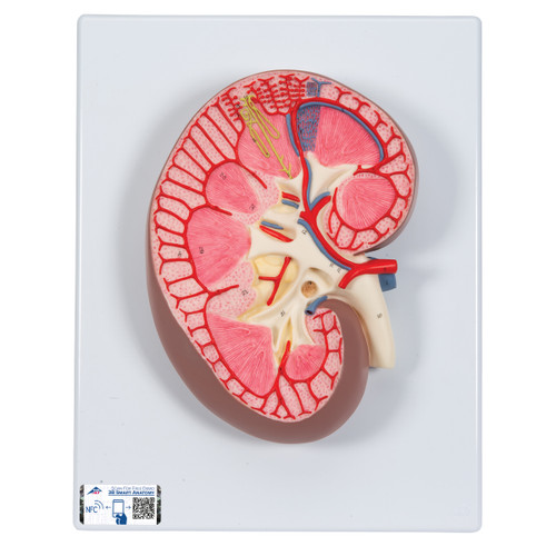
The human nephron model is 120 times life size.
Nephrons and blood vessel model is mounted on a baseboard.
Every original 3B Scientific® Anatomy Model gives you direct access to its digital twin on your smartphone, tablet or desktop device.
Enjoy using the exclusive virtual anatomy content with the following features:
- Freely rotate your digital model and zoom in and out
- Display hotspots and their anatomical structures
- Augmented Reality (AR) feature starts your virtual anatomy model
- Anatomy Quiz function to test and improve your anatomical knowledge with instant results and final score evaluation
- Drawing function that allows image customization with save and share function
- Useful Notes function to help you with your personal learning
- Possibility to learn both male and female anatomy
- Easy access to 3D content both online and offline
- Available in 11 languages
Understanding kidney function begins at the nephron. Enlarged one hundred twenty times, this model makes microanatomy legible from across the room and invites up-close discussion of filtration, reabsorption, and secretion.
A cutaway design lays out the corticomedullary context so you can compare cortical and juxtamedullary nephrons side-by-side. The renal corpuscle shows the capillary tuft (glomerulus) nestled within Bowman’s capsule, with clear afferent and efferent arterioles for explaining hydrostatic pressure and GFR control. Adjacent to the distal convoluted tubule, the juxtaglomerular apparatus (JGA)—including the macula densa—supports conversations about tubuloglomerular feedback and RAAS activation.
The tubular path is easy to follow:
-
PCT with its role in bulk reabsorption
-
Loop of Henle (descending/ascending limbs) illustrating countercurrent mechanics
-
DCT for fine-tuning electrolytes
-
Collecting duct for water balance under ADH influence
Blood-flow pathways are just as explicit. The model depicts peritubular capillaries surrounding cortical segments and vasa recta plunging toward the medulla, perfect for illustrating countercurrent exchange and osmotic gradients that drive concentration of urine.
Use this model to:
-
Walk patients through CKD, hypertension, or diuretic mechanisms
-
Demonstrate how stones, obstruction, or ischemia affect filtration and flow
-
Prep students for exams by tying structure to function (e.g., why the thick ascending limb is a target for loop diuretics)
-
Correlate gross-to-imaging and physiology-to-pharmacology in an integrated lesson
Educational model only. Not intended for diagnosis.
Key Features
-
120× magnification for crystal-clear microanatomy
-
Full nephron pathway: renal corpuscle → PCT → loop of Henle → DCT → collecting duct
-
Microvasculature shown: afferent/efferent arterioles, peritubular capillaries, vasa recta
-
Juxtaglomerular apparatus with macula densa for RAAS and feedback discussions
-
Cortex-to-medulla orientation to explain concentration gradients and urine concentration
-
High-contrast coloring & tactile relief for easy identification in groups
-
Durable, wipe-clean construction on a stable base
-
Includes 3B Smart Anatomy digital content (mobile & web)
What’s Included
-
Nephrons & Blood Vessels Model, 120×, base-mounted
-
Printed reference key (structures & flow overview)
-
3B Smart Anatomy activation (digital learning access)
| Weight | 1.76 lb |
| Dimensions | 10.2 x 7.5 x 2.0 in |
| Brand | 3B Scientific |


