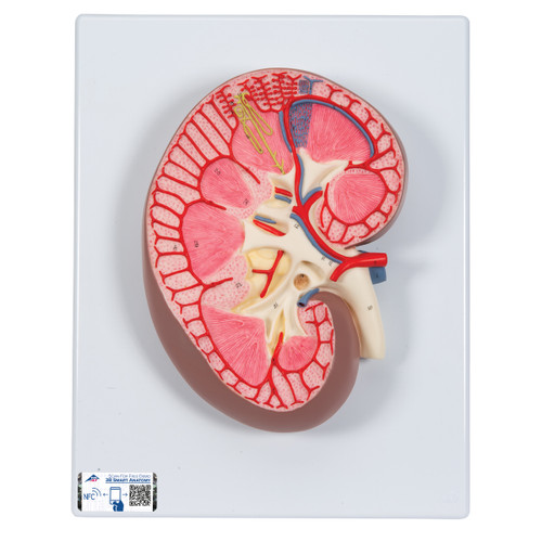
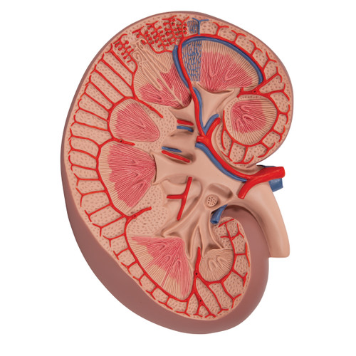
Product details
Your benefits with all 3B Smart Anatomy models:
- Free warranty extension from 3 to 5 years
- Free access to 3B Smart Anatomy training courses in the award-winning Complete Anatomy App
- Includes 11 3B Smart Anatomy training courses with 23 lectures and 117 different interactive virtual model visualisations. Also includes 39 multiple-choice quizzes.
To enjoy all these benefits, simply scan the label and register your 3B Smart Anatomy model online. All features of the 3B Smart Anatomy are available to you completely free of charge.
Learn more about the "Virtual meets Reality" concept developed by 3B Scientific to further enhance the study of human anatomy through the use of all learning tools.
Every original 3B Scientific® Anatomy Model gives you direct access to its digital twin on your smartphone, tablet or desktop device.
Enjoy using the exclusive virtual anatomy content with the following features:
- Freely rotate your digital model and zoom in and out
- Display hotspots and their anatomical structures
- Augmented Reality (AR) feature starts your virtual anatomy model
- Anatomy Quiz function to test and improve your anatomical knowledge with instant results and final score evaluation
- Drawing function that allows image customization with save and share function
- Useful Notes function to help you with your personal learning
- Possibility to learn both male and female anatomy
- Easy access to 3D content both online and offline
- Available in 11 languages
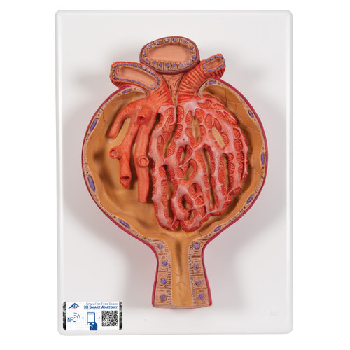
Where kidney function begins, clarity matters. This enlarged model presents the renal corpuscle as a self-contained “learning block,” revealing spatial relationships that are hard to appreciate in slides or diagrams.
-
Bowman’s capsule is shown with distinct parietal and visceral layers. The visceral layer forms podocytes that embrace the capillary tuft, while Bowman’s (urinary) space between the layers illustrates the immediate destination of filtrate.
-
The glomerular capillary tuft is fed by the afferent arteriole and drained by the efferent arteriole at the vascular pole, enabling discussions of pressure, GFR, and autoregulation.
-
The model highlights the tripartite filtration barrier concept—fenestrated endothelium, the glomerular basement membrane (GBM), and podocyte slit diaphragms—for a clean narrative of selectivity and permeability.
-
Mesangial regions are depicted to support conversation on structural support, flow regulation, and immune complex handling.
-
At the urinary pole, Bowman’s space communicates with the beginning of the proximal convoluted tubule (PCT), providing a natural segue into reabsorption and the nephron continuum.
-
An adjacent juxtaglomerular apparatus (JGA) zone (macula densa region and JG cells) is represented schematically to connect corpuscle anatomy with tubuloglomerular feedback and RAAS concepts.
Use this model to:
-
Explain how filtrate forms and why BUN/creatinine rise when the barrier or pressures change
-
Show where albumin is retained (and why albuminuria signals barrier damage)
-
Demonstrate sites of injury in glomerulonephritides and diabetic nephropathy
-
Teach autoregulation (afferent vs. efferent tone) and the impact of drugs (e.g., ACE inhibitors, NSAIDs)
-
Bridge microscopy with clinical imaging and lab interpretation
Educational model only. Not intended for diagnosis.
Key Features
-
700× magnification of the renal corpuscle for group teaching
-
Clear depiction of glomerulus, Bowman’s capsule, Bowman’s space, afferent/efferent arterioles
-
Emphasizes the filtration barrier (endothelium → GBM → podocyte slits)
-
Shows vascular pole and urinary pole with transition to PCT
-
Mesangial regions and JGA context for physiology integration
-
High-contrast coloring & tactile relief to speed identification
-
Rigid, wipe-clean build on a stable base; designed for daily use
-
Includes 3B Smart Anatomy access (mobile & web) for extended learning
What’s Included
-
Model of Malpighian (Renal) Corpuscle, 700×, base-mounted
-
Printed reference/overview key (structures & concepts)
-
3B Smart Anatomy activation (digital content access)
Specifications (Approx.)
-
Magnification: 700× microanatomy
-
Structures shown: Glomerular capillaries, Bowman’s capsule (parietal/visceral), Bowman’s space; afferent/efferent arterioles (vascular pole); urinary pole with PCT origin; mesangial areas; JGA context (macula densa/JG cells schematic)
-
Format: Desktop, base-mounted educational model
-
Materials: Rigid polymer with matte, glare-reduced finish
-
Program: 3B Smart Anatomy
-
SKU: 005-1000298
(Exact dimensions/weights may vary slightly by production lot.)
Who It’s For
-
Nephrology, urology, dialysis clinics (patient counseling)
-
Medical, nursing, PA, and allied-health programs (anatomy/physiology)
-
Pharmacy education (drug mechanisms: ACEi/ARB, NSAIDs, SGLT2 context)
-
Public health educators (kidney screening & awareness)
| Weight | 1.61 lb |
| Dimensions | 10.2 x 7.5 x 3.1 in |
| Brand | 3B Scientific |

