Product details
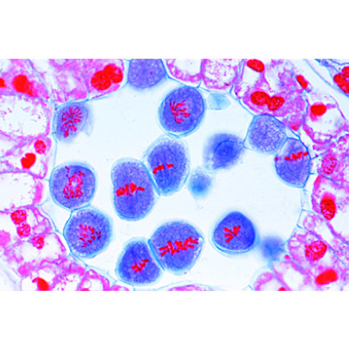
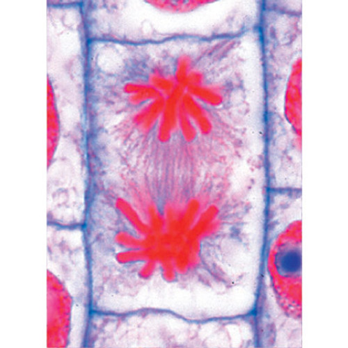
· Chromosomes coloured according to modified AZAN staining colours
· Cell components are colour-coded in accordance with educational aspects
· Attaching magnets on the rear
· Storage system, free-standing or hanging up.
· Supplied with detailed description and copying templates
· Enlarged 10,000 times
The three-dimensional relief model shows the 10 stages of meiosis on the basis of a typical mammal cell:
1. Interphase (stage of G1-phase)
2. Prophase I (leptotene)
3. Prophase I (zygotene and pachytene)
4. Prophase I (diplotene)
5. Prophase I (diakinesis)
6. Metaphase I
7. Anaphase I
8. Telophase I, cytokinesis I, interkinesis, prophase II and metaphase II
9. Anaphase II
10. Telophase II and cytokinesis II
Dimensions: approx. 60x40x6 cm³
Weight: approx. 1.7 kg
The Meiosis Model (005-1013869) is an educational tool designed to demonstrate the complex process of cell division that leads to the formation of gametes. This model clearly illustrates the phases of meiosis I and meiosis II, including prophase, metaphase, anaphase, and telophase, as well as crossing-over and reduction division.
Ideal for biology classrooms, laboratories, and advanced science courses, the model helps students understand genetic variation, chromosome behavior, and the differences between mitosis and meiosis. It is particularly valuable for teaching genetics, reproductive biology, and life sciences, making abstract cellular processes easier to visualize and explain.
Durable and precisely crafted, the Meiosis Model is a reliable teaching aid for enhancing lessons, supporting laboratory studies, and engaging students in hands-on learning.
Documentation
| Weight | 1.904 kg |
| Dimensions | 60 x 40 x 6 cm |
| Brand | 3B Scientific |


