Product details
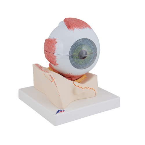
The human eyeball can be dissected into:
- Both halves of sclera with cornea and eye muscle attachments
- Both halves of the choroid with iris and retina
- Eye lens
- Vitreous humour
Every original 3B Scientific® Anatomy Model gives you direct access to its digital twin on your smartphone, tablet or desktop device.
Enjoy using the exclusive virtual anatomy content with the following features:
- Freely rotate your digital model and zoom in and out
- Display hotspots and their anatomical structures
- Augmented Reality (AR) feature starts your virtual anatomy model
- Anatomy Quiz function to test and improve your anatomical knowledge with instant results and final score evaluation
- Drawing function that allows image customization with save and share function
- Useful Notes function to help you with your personal learning
- Possibility to learn both male and female anatomy
- Easy access to 3D content both online and offline
- Available in 11 languages
The Human Eye Model, 3 times full-size, 7 part (005-1000258) offers a clear and accurate representation of the external and internal anatomy of the human eye. Enlarged to three times life-size, this model provides excellent visibility while remaining compact enough for classroom and clinical use.
It separates into seven removable components, including the sclera with choroid and retina, cornea, iris, lens, vitreous body, and optic nerve, making it easy to study each structure in detail. This hands-on dissection capability makes the model especially useful for teaching ophthalmology, optometry, anatomy, and patient education, where understanding the eye’s anatomy is essential.
Durable and designed for repeated use, this 3B Smart Anatomy eye model is ideal for universities, medical training centers, and healthcare professionals who need a reliable and visually clear tool for demonstrating eye function and vision-related conditions.
Documentation
| Weight | 2.98 lb |
| Dimensions | 7.1 x 10.2 x 7.5 in |
| Brand | 3B Scientific |

