Product details
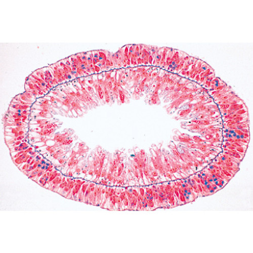
1(e). Hydra, fresh water polyp, t.s. with ectoderm and entoderm
2(d). Carabus, ground beetle, gizzard
3(c). Salivary gland of cat, t.s.
4(c). Esophagus of cat, t.s.
5(d). Fundic stomach of cat, t.s.
6(c). Small intestine of cat, t.s. routine stained
7(f). Small intestine, t.s. blood vessels injected
8(d). Appendix of human, t.s.
9(c). Large intestine of cat, t.s.
10(c). Liver of pig, t.s.
11(f). Malpighian tubules of insect, t.s.
12(c). Primordial kidney (mesonephros) of frog, t.s.
13(d). Hind-kidney (metanephros) of rabbit, t.s.
14(d). Kidney of mouse with pelvis, l.s.
15(f). Kidney of mouse, t.s. injected to show storage.
| Weight | 0.22 kg |
| Brand | LIEDER |
The Series II. Metabolism – Slides set offers a detailed look into the microscopic processes and structures involved in biological metabolism. This collection includes expertly prepared slides that demonstrate key aspects of enzyme activity, energy transformation, and cellular processes such as photosynthesis and respiration.
Designed for teaching biology, biochemistry, and life sciences, this set allows students to visually connect theoretical knowledge with practical examples. Each slide is professionally prepared to ensure clarity and durability, making them ideal for repeated use in classrooms, universities, and laboratories. Together with other series sets, it provides a structured foundation for understanding essential life processes.


