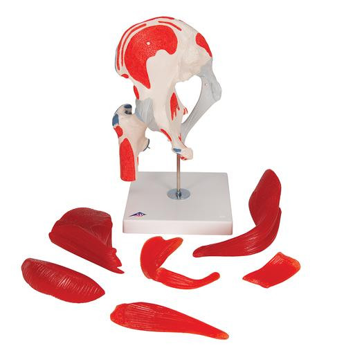
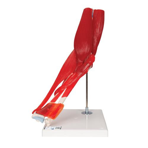
The model consists of 12 parts which can be easily disassembled for easy demonstration. The muscles and muscle portions can be taken off, and a special color coding has been used to show muscle origin and insertion points on the femur, tibia, and fibula. Additionally, the model also shows parts of the fibular and tibial collateral ligaments.
Whether your are studying or teaching the deeper anatomical layers of the human knee joint, or are looking for a tool to better educate your patients about their injuries and rehab, the Knee Joint Model by 3B Scientific® is an extraordinary teaching tool and offers exceptional value.
Every original 3B Scientific® Anatomy Model gives you direct access to its digital twin on your smartphone, tablet or desktop device.
Enjoy using the exclusive virtual anatomy content with the following features:
- Freely rotate your digital model and zoom in and out
- Display hotspots and their anatomical structures
- Augmented Reality (AR) feature starts your virtual anatomy model
- Anatomy Quiz function to test and improve your anatomical knowledge with instant results and final score evaluation
- Drawing function that allows image customization with save and share function
- Useful Notes function to help you with your personal learning
- Possibility to learn both male and female anatomy
- Easy access to 3D content both online and offline
- Available in 11 languages
To get started, simply scan the QR-code located on your 3B Scientific® Anatomical Model, download the new 3B Smart Anatomy app and step into the virtual world of Human Anatomy. Click here to learn more.
Documentation
| Weight | 3.86 lb |
| Dimensions | 13.0 x 6.7 x 6.7 in |
| Brand | 3B Scientific |


